Movement is life. This trite statement takes on a profound, often painful meaning when that movement is taken away from us. When every step becomes a challenge, when raising a hand to comb our hair causes a grimace of pain, or when an emergency takes us out of activity for months - then we enter the world of orthopaedics.
The human body is a masterpiece of bioengineering. A skeleton made up of more than 200 bones, connected by a system of joints, ligaments and tendons, and then powered by hundreds of muscles controlled by the nervous system, allows us to do extraordinary things. We can run marathons, thread a needle with precision, lift weights and perform acrobatics. However, this complex machinery, like any other, is subject to failure. Sometimes it is a sudden mechanical failure (injury) and sometimes it is slow wear and tear of the material (degeneration).
Orthopaedics is the branch of medicine that has taken on the task of fixing the machine. For years associated only with „setting bones” and plastering, today it is one of the most technologically advanced specialities, benefiting from robotics, materials engineering and molecular biology.
This monumental guide will take you on a journey through the fascinating world of the musculoskeletal system. You'll understand how your joints work, why your back hurts, how doctors diagnose and modern methods of restoring function.
What is Orthopaedics really? (Historical and Contemporary Perspectives)
The word „orthopaedics” is derived from the Greek: orthos means „simple” and paidion - „child”. The term was coined in 1741 by Nicolas Andry when he published a book on correcting deformities in children. Initially, therefore, orthopaedics was concerned with straightening spines and limbs in the youngest children (hence the symbol of the „tree linked to a post”).
Today, the definition is much broader. Modern musculoskeletal orthopaedics and traumatology deals with the diagnosis, treatment (conservative and surgical) and prevention of diseases of the entire skeleton (except the cranial bones), the ligamentous-articular system and the muscular system.
It is an interdisciplinary field. An orthopaedic surgeon has to be a bit of a radiologist (to read complex images), a bit of a neurologist (to distinguish between root and joint pain) and, above all, an excellent mechanic of the human body.
Functional Anatomy: Understanding the Machine
To understand disease, we must first understand physiology. Our musculoskeletal system is an interconnected system of vessels. The failure of one component (e.g. the foot) entails an avalanche of changes elsewhere (knee, hip, spine).
1. bones - scaffolding
It is the hardest tissue, but not at all dead. Bone is a living, dynamic organ that undergoes remodelling (remodelling) throughout life. Osteoblasts build bone, osteoclasts destroy it. This allows bones to knit together after złamanias and adapt to stresses.
2. joints - Hinges
Bone junction sites. The most important from an orthopaedic point of view are the synovial joints (e.g. knee, hip). Their ends are covered by vitreous cartilage - an incredibly smooth, elastic tissue that minimises friction. The whole is enclosed in a joint capsule filled with synovial fluid („grease”). This cartilage damage is the cause of most orthopaedic pain.
3. ligaments and tendons - cables and ropes
Often confused by patients.
- Ligaments: They connect bone to bone. They are like bands that stabilise the joint. When they break (e.g. the ACL in the knee), the joint becomes unstable and „runs away”.
- Tendons: They connect the muscle to the bone. They transmit the force of the muscle to the skeleton, setting it in motion. The most famous are the Achilles tendon.
4 Muscle - Engine
The active part of the musculoskeletal system. Without them, the skeleton would just be a pile of bones. Muscles not only move the body, but also act as „dynamic ligaments”, stabilising joints. Strong muscles are the best protection against injury.
Orthopaedic Diagnostics: The Art of Detection
A visit to an orthopaedic surgeon is more than looking at an X-ray. It is an investigative process.
Interview: Key to the Riddle
The doctor asks: „When does it hurt?”. Pain in the morning that passes after pacing suggests degenerative or inflammatory changes. Pain that occurs after exercise and increases in the evening is often overload. Sudden, sharp pain, accompanied by a snap, is an injury. Every detail matters.
Physical Examination: Hands that see
This is the most important moment of the visit. The orthopaedic surgeon does not examine the „photo”, he examines the patient. He performs a series of functional tests:
- Meniscus tests (twisting the knee to cause pain or „clicking”).
- Resistance tests (the patient wrestles with the doctor, which allows the strength of the muscles and the condition of the tendons to be assessed).
- Range of motion test (Does the hand rise to the end?).
- Stability test (Doesn't the knee „scoff”?).
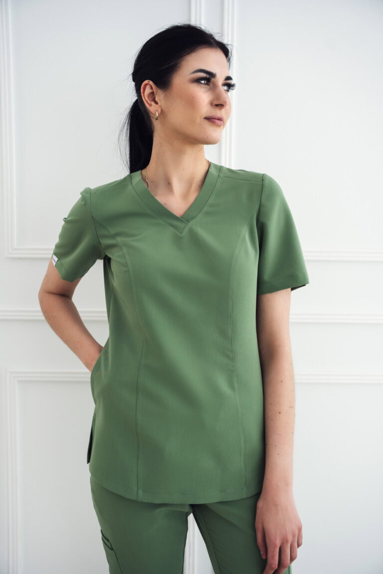
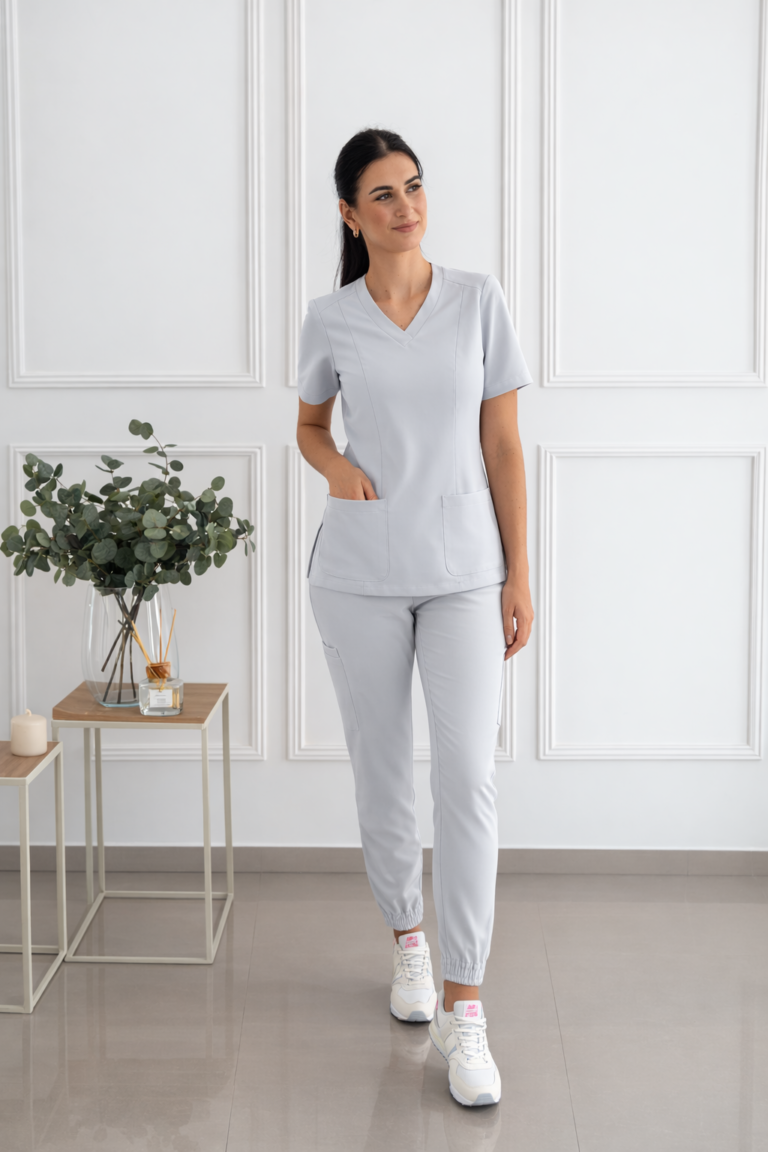
This examination requires the doctor to be very physically fit. The orthopaedic surgeon squats, bends, lifts the patient's limbs (often inert and heavy), exerts force and resistance many times during the day. This is physical work. This is why rigid aprons are a thing of the past in modern orthopaedics. The standard has become ergonomic medical suit. The doctor, when performing the precise Lachman test (for the cruciate ligament), must have full freedom of movement of the arms and trunk. Flexible medical uniforms of modern fabrics allow him to focus on the diagnosis rather than struggling with uncomfortable clothing. This is particularly evident in paediatric or sports orthopaedists, where the dynamics of the examination are greatest.
Imaging Diagnostics: Insights from the Inside
Only after the manual examination comes the technology:
- X-ray (X-ray): Base. Shows bones, złamania, degeneration, bone spurs. Does not show soft tissues!
- Ultrasonography (USG): „Orthopaedic stethoscope. Allows live assessment of muscles, tendons, ligaments, joint effusion. The doctor can see how the tissues behave in motion.
- MRI (Magnetic Resonance Imaging): Gold standard. It shows everything with amazing precision - meniscus, cartilage, bone marrow swelling, discs in the spine.
- CT (computed tomography): Used mainly for complex złamania to plan the operation (3D bone modelling).
The Big Four: The Most Common Orthopaedic Problems
What do we most often come to the surgery with? The spectrum is huge, but certain conditions are the scourges of our time.
1. Degenerative joint disease (Osteoarthrosis)
It is the price we pay for longevity and... evolution. It most commonly affects the knees (gonarthrosis) and hips (coxarthrosis).
- Mechanism: The articular cartilage, which has no blood supply and innervation of its own, slowly wears away. It loses its smoothness and becomes rough. The bone beneath the cartilage begins to „feel” the load, reacting with pain and the formation of bone outgrowths (osteophytes). The joint loses its glide.
- Symptoms: Startle pain (on standing up), morning stiffness, crackling in the joint, restriction of mobility (difficulty putting on a sock with a bad hip).
2 Spinal Pain: A Civilisation Epidemic
The spine is our axis, but structurally it is not ideally suited to sitting for eight hours a day.
- Discopathy: The intervertebral disc (disk) is a shock absorber. With age or from overload, it loses water, flattens out and its nucleus pulposus can slip out (herniation). If it presses on the nerve root, we have sciatica or shoulder pain - pain radiating into the leg or arm, often with numbness and weakness.
- Overload changes: Often, back pain does not come from a disc, but from muscles and fascias that are permanently strained due to stress and złhis position.
3 Sports Injuries: The Price of Ambition
Physical activity is health, but sport (especially amateur sport without preparation) is a frequent client of orthopaedics.
- Rupture of the ACL (Anterior Cruciate Ligament): A nightmare for skiers and footballers. Sudden twisting of the knee, cracking, swelling and instability („running away” of the knee).
- Meniscus injuries: The menisci are shock absorbers in the knee. They rupture with deep squats or rotation. A locked meniscus can immobilise the knee.
- Tennis/golfers' elbow: It is not a problem of the joint, but of the tendon attachment. Pain when gripping a cup or saying hello. It is due to microtrauma and lack of regeneration.
- Ankle sprain: The popular „ankle sprain”. Often underestimated, it leads to chronic instability and recurrent injury.
4. carpal tunnel syndrome
Office sickness. Working with a mouse and keyboard causes swelling in the carpal tunnel, compressing the median nerve. Symptoms include numbness in the thumb, index and middle finger (especially at night), and objects falling out of the hands.
Paediatric Orthopaedics: Not Just a Small Adult
Children are a specific group of patients. Their bones are soft, flexible but have growth zones (growth cartilage). Damage to such a zone can inhibit limb growth.
The most common problems in the youngest are:
- Hip dysplasia: Acetabular joint underdevelopment in infants. Thanks to common hip ultrasound examinations, this problem is nowadays detected quickly and treated non-invasively.
- Scoliosis: Tri-plane curvature of the spine. It is not just a „crooked back”, it is a rotation of the vertebrae that can deform the rib cage and compress the lungs. Detected early (during the growth spurt), it can be corrected with a corset and rehabilitation.
- Foot defects: Flat feet, clubfoot. Require differentiation between physiology (children naturally have flat feet up to a certain age) and pathology requiring orthotics or surgery.
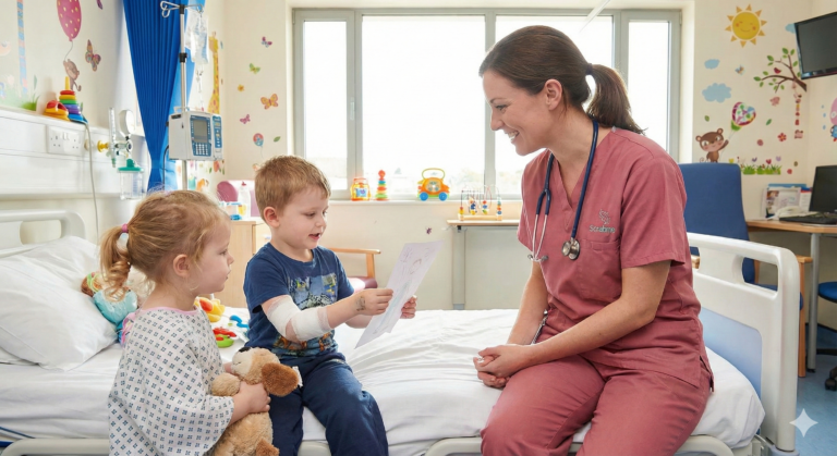
Working with children requires the orthopaedic surgeon to have not only knowledge but also a psychological approach. Children's surgeries are increasingly moving away from the fear-inducing „white coat” („white coat syndrome”). Colourful, friendly medical scrubs for women and masculine, in shades such as mint, pink or blue, help to shorten the distance. A child, seeing a doctor dressed in „colourful” clothes, is less afraid of the examination, which makes it easier to make a diagnosis.
Traumatology: When Seconds Matter
Traumatology (trauma surgery) is the sister of orthopaedics. It deals with sudden injuries: 1TP1Fractures, dislocations, multi-organ injuries after traffic accidents.
Here, the action is different. There is no time for weeks of planning. An open fracture, hip dislocation or 1TP1Pelvic fracture are medical emergencies and sometimes life-threatening. The traumatologist is a chaos engineer. He has to put together a „jigsaw puzzle” of bones, using titanium plates, screws, intramedullary nails and external stabilisers. The aim is not only to fuse the bone, but to restore its axis, length and function.
Modern traumatology strives for a so-called stable anastomosis, which allows movement almost immediately after surgery. The times when a patient with a złamana thigh lay for three months on a lift are gone. Today, after a nail anastomosis, the patient gets out of bed on the second day. This prevents embolic complications and muscle atrophy.
The Hand - Excellent and Fragile Tool
Hand surgery is actually a separate sub-speciality, combining orthopaedics, plastic and vascular surgery. The hand is a mechanism of incredible złożonicity - 27 bones, hundreds of ligaments, thread-thick nerves. Orthopaedic surgeons who deal with the hand treat:
- Dupuytren's contracture: Scarring of the fascia of the hand, which pinches the fingers permanently to the inside of the hand.
- Finger snapping: Inflammation of the tendon sheath that blocks the finger in flexion.
- Tendon and nerve injuries: They require microsurgery - suturing under a microscope, using threads thinner than a human hair.
This is a field that requires incredible precision, patience and... ergonomics. Many hours of microsurgical operations in a forced position require the surgeon to be in absolute thermal and movement comfort, which is provided by modern medical textiles.
Engineering the Human Body: The Complete Guide to Orthopaedics. Part 2: Treatment, Surgery and the Road to Fitness.
In the first part of our guide (which you can find here: [LINK TO PART 1]), we learned about the anatomy of the musculoskeletal system and the most common malfunctions it can suffer. We already know how an orthopaedic surgeon makes a diagnosis. Now it's time for the most important question: how to fix it?
Modern orthopaedics is a fascinating array of possibilities. On the one hand, we have powerful biological engineering (stem cells, growth factors), on the other, precision mechanics (titanium implants, surgical robots). However, at the centre of this process is always the c1TP1Person - the patient struggling to recover - and the medical team, whose work is one of the greatest physical challenges in the medical world.
In this section, we'll step into the operating theatre, look at modern technology, understand the role of rehabilitation and why, for the orthopaedic surgeon, its medical suit is as important as the scalpel.
Chapter 5: Recovery Strategy - Behavioural Treatment (Before We Use the Scalpel)
Contrary to popular fear, a visit to an orthopaedic surgeon rarely results in a referral to the operating table. The golden rule is: surgery is a last resort. First, we use the body's natural ability to regenerate.
1. Targeted pharmacotherapy
Pain prevents rehabilitation and inflammation destroys tissues. Therefore, treatment starts with NSAIDs (Non-steroidal anti-inflammatory drugs).
- Locks: Precise administration of a steroid to the site of pain. They give quick relief but do not treat the cause. They are an „extinguisher” for an acute inflammatory fire.
- Viscosupplementation: „Joint lubrication”. Hyaluronic acid is injected into the joint (usually the knee). This acts as a shock absorber and glide, reducing the friction of damaged surfaces.
2. Biological Orthopaedics (Regenerative Medicine)
It's a przyszłity that is happening today. Instead of artificial drugs, we use the patient's blood.
- PRP (Platelet Rich Plasma): The patient's blood is drawn, centrifuged and a platelet concentrate is obtained. These platelets release powerful growth factors that stimulate the healing of tendons (e.g. tennis elbow), muscles and ligaments.
- Stem Cells: Taken from bone marrow or fat, they have the potential to transform into other tissues and strongly inhibit inflammation.
3 Physiotherapy - A Real Hero
Without a physiotherapist, the orthopaedist is helpless. Manual therapy, deep tissue massage, dry needling and stabilisation exercises are the key to success in back pain or strains. Often „fixing” złhese movement habits are enough to avoid surgery.
Chapter 6: The 21st Century Operating Room - Minimally Invasive Surgery
If conservative methods fail, surgery steps in. But today's orthopaedic surgery is radically different from that of 20 years ago. We aim to minimise the injury.
Arthroscopy - Keyhole surgery„
It's a revolution. Instead of opening the whole knee or shoulder (which involves pain, scarring and long healing), the surgeon makes two small incisions (about 1 cm).
- A camera (arthroscope) with a cold light is inserted through one.
- Through the second, he introduces miniature tools.
On a monitor screen, in 4K magnification, the doctor can see the inside of the joint. He or she can suture a ruptured meniscus, smooth out cartilage (shaving) or reconstruct a torn ligament using tendons taken from the patient. The patient often goes home the next day.
Osteotomy - Correction of the Axis
Before replacing the joint with an artificial one, you can try to save it by changing the geometry of the bones. If you have a crooked leg (talus/bow), you only put weight on one side of the knee. An osteotomy involves cutting through the bone and putting it in a new, correct axis. This „buys time” for your own joint.
Chapter 7: The Final Solution - Endoprosthesis (Joint Replacement)
When a joint is so damaged that bone rubs against bone and pain wakes you up at night - the only salvation is to replace the parts with new ones.
How does it work?
The surgeon removes the damaged joint surfaces and replaces them with implants made of titanium, ceramic and highly cross-linked polyethylene.
- Hip Endoprosthesis: It is the most common and successful operation in orthopaedics. It restores patients who have been in a wheelchair to the ability to walk and even play sports.
- Knee Endoprosthesis: More complicated because the knee is not a simple hinge. Modern implants mimic natural mobility.
These are „Quality of Life” operations - restoring the joy of life.
Chapter 8: Orthopaedics is Physical Work - The Role of the Team and Ergonomics
When we watch medical series, we see surgeons in clean, spotless outfits. The reality of the orthopaedic operating theatre is different. It is hard physical work.
Orthopaedics is „carpentry” on a living organism. Oscillating saws, drills, hammers, chisels and screwdrivers are used. Replacing a hip joint in an upright man requires the surgeon to use a great deal of physical force - when setting dislocations, driving in the endoprosthesis stem or reaming (reaming) the acetabulum.
Extreme conditions
The operating theatre is sterile. The surgeon is dressed in an impermeable, sterile apron, often wearing a helmet with a ventilation system on his head (to protect against infection) and wearing a heavy lead apron (to protect against X-rays, which are used to view the bones). Added to this is the stress and heat of the operating lamps. The temperature under the clothes rises rapidly.
Under such conditions, what the doctor has underneath - i.e. his medical suit - is becoming a key element of health and safety.
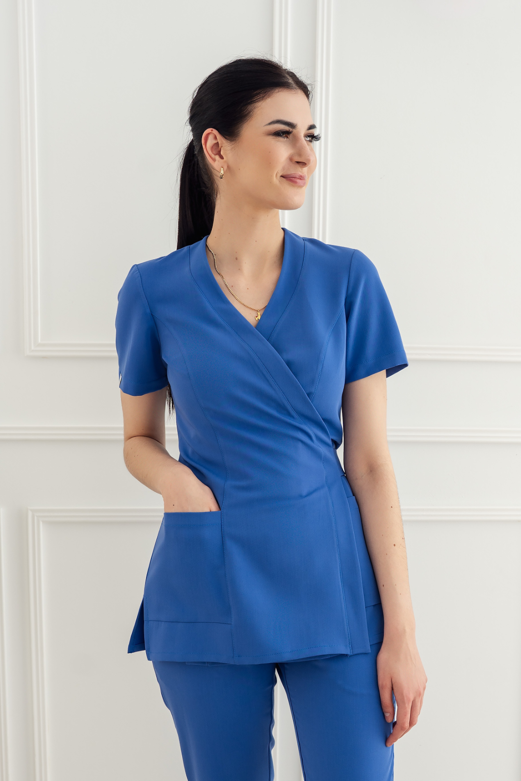
- Thermoregulation: A plain cotton T-shirt under a sterile apron is a guarantee of soaking wet from sweat after 30 minutes. Modern medical uniforms (scrubs) made from polyester and viscose blends act like sportswear - they wick moisture away from the body. This allows the surgeon to remain comfortable and focused for the 3-4 hours of the procedure.
- Freedom of Movement: The orthopaedist operates with the whole body. He has to wrestle with his legs, use arm strength. If his trousers restrain him, he loses stability. Flexible medical trousers (often with added elastane) are essential here.
- Role of the Team: Standing next to the surgeon are the instrumentalists and anaesthetists. For them, too, comfort is crucial. The instrument attendant stands still for hours, administering hundreds of instruments. Comfortable medical scrubs for women with an ergonomic cut to reduce static fatigue.
W Scrabme We understand this very well. We know that there is no room for discomfort in the orthopaedic block. Clothing has to be a second skin that you forget about so that you can focus solely on the patient.
Chapter 9: Rehabilitation - 50% of Success
The operation is only halfway. The best surgery, performed by a professor, will not succeed without rehabilitation.
After surgery (e.g. ligament reconstruction), the muscles atrophy at an express rate. The joint stiffens. Rehabilitation is painstaking work to regain range of movement, strength and deep sensation (proprioception).
- The „Motion is Lotion” principle: Movement is like a balm for the joint. It nourishes the cartilage. This is why patients are often „uprighted” (put on their feet) as early as the first day after surgery.
Chapter 10: Prevention - How Not to Become an Orthopaedic Patient?
Can a visit to the orthopaedist be avoided? In many cases, yes. Our musculoskeletal system is damaged by two things: stillness i overloads.
1. Work Ergonomics
If you work at a desk, your spine suffers. Make sure you have a monitor at eye level, a good chair and - most importantly - movement breaks every hour. If you work physically (e.g. as a nurse or physiotherapist), learn safe patient lifting techniques (using your legs, not your back!). This is also where appropriate, non-restrictive clothing helps.
2. Weight Control
It is simple physics. Every extra kilogram of body weight is approximately 4-5 kg of extra load on the knees when walking up stairs. Obesity is a major joint killer. Losing 5kg can reduce knee pain by 50%.
3 Wise Motion
„Sport is health”, but only sport practised with a head. Weekend warriors who sit for a week and run 10km on Saturday are asking for injury. Regularity and warm-ups are key.
4 Footwear
The feet are the foundation. Bad shoes (stilettos, flat ballerinas without support) change the alignment of the entire body axis, damaging the knees and spine.
Summary: Movement is Freedom
Orthopaedics is a beautiful field. It is medicine that restores freedom. Freedom to move without pain, freedom to return to work, to passions, to play with children.
From the precise diagnosis, to the advanced surgical technology, to the sweat poured in the rehabilitation room - it is a team process. A process where technology meets biology and the physical strength of the doctor meets the delicacy of the tissue.
Take care of your musculoskeletal system - it's the only one you have. But if it refuses to obey, know that modern orthopaedics has the tools to get you back on your feet.
Joint pain restricting your life? Don't wait for it to „go away on its own”. Consult an orthopaedic specialist and regain the joy of movement. Time is against you.
Are you an orthopaedic surgeon, an instrumentalist, a physiotherapist? Your job is physically challenging. Don't let your outfit be an extra burden. Choose a professional Scrabme medical clothing. Our medical uniforms have been tested under the toughest conditions to give you the thermoregulation and freedom you need at the operating table.